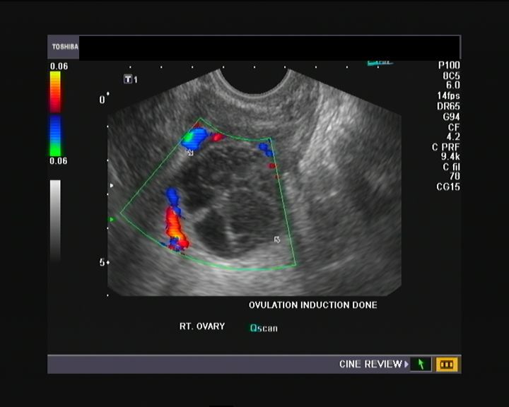
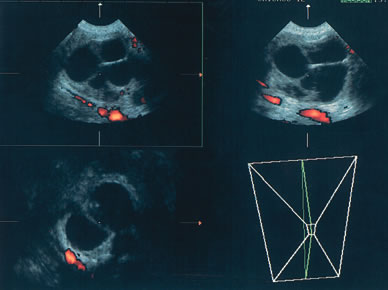
Early Stage Ovarian Cancer Color Normal Ovary Doppler Ultrasound

Selection Of Hens With Normal Ovaries Or Ovarian Tumors Using A C Download Scientific Diagram

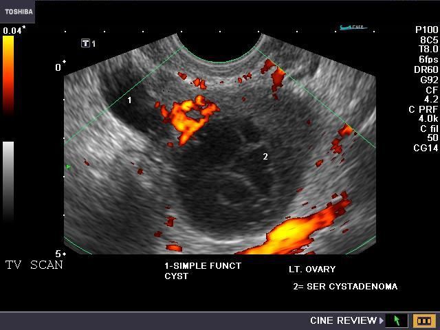
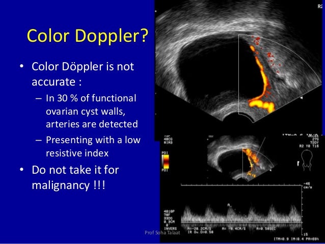
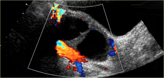
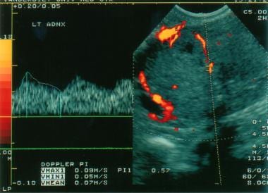
Ovarian Cancer Color Doppler

Ultrasonographic Color Doppler Appearance Of The Ovaries With Moderate Download Scientific Diagram

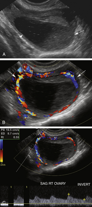
Color Doppler Image Of The Right Ovary Demonstrates The Typical Ring Download Scientific Diagram
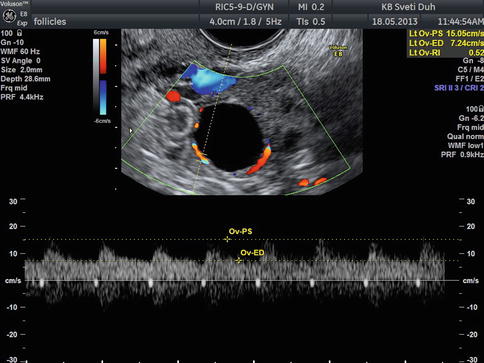
A Gallery Of High Resolution Ultrasound Color Doppler 3d Images Ovaries
A Gallery Of High Resolution Ultrasound Color Doppler 3d Images Ovarian Masses

A small sample is sent to a laboratory where a pathologist examines it under a microscope.
Early stage ovarian cancer color normal ovary doppler ultrasound.
Duplex Ultrasound Evaluation Of The Uterus And Ovaries Radiology Key
The Normal Ovary Changes In The Menstrual Cycle Radiology Key

Detection Of Ovarian Tumors In Chicken By Sonography Barua 2007 Journal Of Ultrasound In Medicine Wiley Online Library
Doppler In Gyneacology Dr Muhammad Bin Zulfiqar

Iame Sonography Of The Ovary Benign Vs Malignant
The Radiology Assistant Roadmap To Evaluate Ovarian Cysts
Https Onlinelibrary Wiley Com Doi Pdf 10 7863 Jum 1992 11 12 631
Ultrasound Imaging Of Ovarian Cancer Chapter 22 Ultrasonography In Gynecology

Emergency Ultrasound
Imaging Techniques In Gynaecology Obgyn Key
Diagnostic Ultrasound In The Assessment Of The Adnexal Mass Glowm
Https Pubs Rsna Org Doi Pdf 10 1148 Rg 285075130

Color Flow Doppler Evaluation Of Uterus And Ovaries And Its Optimization Techniques Sciencedirect
Https Obgyn Onlinelibrary Wiley Com Doi Pdf 10 1002 Uog 5365

Wk 3 L 2 Figure 2 Transvaginal Color Doppler From A Cervical Carcinoma A Highly Vascularized Area Is Clearly Seen Wit Cervical Carcinoma Myelofibrosis Cervix

Selected Images From A First Trimester Ultrasound There Is A Hypoechoic Centrally Cystic Cyst In The Left Ovary Corpus Luteum Polycystic Ovaries Ovarian Cyst

Ultrasound Doppler Principles Preparation Results And More

Pdf Transvaginal Ultrasound Assessment Of The Premenopausal Ovarian Mass
Https Encrypted Tbn0 Gstatic Com Images Q Tbn 3aand9gcs7vpb9rgiygvimx Cdktgsyo2hdplbbiiz7ewvyyuxhpcu92mc Usqp Cau

Diagnosis And Treatment Of Uterine And Pelvic Arteriovenous Malformations Endovascular Today

Differences In The Angiogenesis Of Benign And Malignant Ovarian Tumors Demonstrated By Analyses Of Color Doppler Ultrasound Immunohistochemistry And Microvessel Density Emoto 1997 Cancer Wiley Online Library
Which Ultrasound Findings Are Characteristic Of Malignant Ovarian Tumors

Full Text Assessment Of Adnexal Masses Using Ultrasound A Practical Review Ijwh
Can Ovaries Be Preserved After An Ovarian Arteriovenous Disconnection One Case Report And A Review Of Surgical Treatment Using Da Vinci Robots For Aggressive Ovarian Fibromatosis Springerlink
Source : pinterest.com
